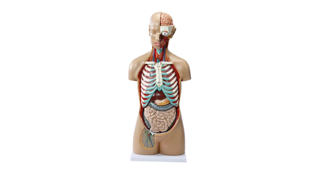
| Highly detailed torso with 16 removable parts, open back and genderless |
3rd display in the range:
1) Price : DKK 2,825 (incl. VAT)
2) Size : 85 cm in height and weighs approximately 9 kg
3) Removable parts : 16 pcs.
4) Color, stand, material quality and inclusive/exclusive list of names
The skin looks like rubber, which shines. The other colors appear muted, slightly dark and shiny. The stand is an approximately 2.3 cm thick plastic plate in grey-white, which measures approx. 23 x 33 cm.
The material quality seems good and very robust in all the model's tissues, although the skin and especially some tissues look very artificial (e.g. the eye and all vertebrae except the 7th thoracic vertebra).
On the torso model, the most important anatomical structures are numbered. An English name list is included.
5) Anatomical details (mentioned from the top of the model downwards in the following 4 groups)
I - The musculoskeletal system (bones, cartilage, ligaments, fascia and skeletal muscles)
Bones : There are quite a few - mainly in the head, chest and back. In the head, the left part of the skull can be seen inside with very few details such as protrusions and holes for vessels and nerves. Externally, a bit of the facial skeleton can be seen around the left eye, where the various bones are seen without details such as protrusions, holes, etc. Furthermore, an insignificant small part of the lower jaw on the right side and the tongue bone can be seen without significant muscle attachments.
Inside the torso, the clavicles and ribs can be seen in cross-section at the open and transected chest wall, when half of each lung has been removed. When the lungs have been removed, the anterior ribs, cartilage and part of the sternum, which is attached to the lungs, can also be seen. This bone tissue is also not seen in detail - however, the anguli sterni is seen. In addition, the iliac crests, quite a bit of the pubic bones and the transition between the 5th lumbar vertebra and the sacrum in the pelvis are visible.
The open back shows different parts of the vertebrae from the upper neck joint to the sacrum (it is difficult to see the coccyx). Pins are not included. The vertebrae are thus "open", so that the vertebral canal with nerves is visible. These bones are seen in the color beige, and virtually no detail is seen in the bone tissue (but studs are seen). The 7th thoracic vertebra, on the other hand, is complete and completely different. It can be taken out. It looks like a real vertebra, is white and the quality is good with many details such as tiny bony projections etc. When this vertebra is taken out, a disc can be seen, which is just a painted illustration in 2 different colors. In a large part of the back, the facet joints are also visible, which are very artificial.
Cartilage is shown in green color on the nose, in the throat, in the trachea, in the chest (rib cartilage) and at the 5th lumbar vertebra in the torso (disc). Ligaments virtually invisible. Fascia not seen either.
Skeletal muscles seen to a lesser extent. In the head, some muscles can be seen around the left side of the mouth and jaw and at the right cheek. In addition, few pectoral muscles, few abdominal muscles and few muscles in the pelvis can be identified. In the open back, musculature can be seen under the skin and between the vertebrae. Apart from a few, it is generally difficult to identify specific muscles in the back.
II – Blood vessels (arteries and veins), nerves and the lymphatic system as well as other things such as salivary glands
Both the very largest arteries and veins as well as quite a few smaller blood vessels are included. In addition to the large vessels in the neck, chest and stomach, detailed blood supply (arteries and veins) can also be seen in many other organs. As for the brain, arteries can be seen on the entire surface of the left hemisphere (which can be taken out), the internal carotid artery, the superior and inferior sagittal sinuses and a bit of the circulus arteriosus cerebri at the base of the skull (on the left side).
Smaller blood vessels are also seen in the heart ("coronary vessels"), the lungs, the stomach, the small and large intestine, the pancreas, the liver and the right kidney (which can be opened). Openings in the surface of the spleen and right thigh also show some blood vessels. In the back, veins and the vertebral artery can be seen.
The nervous system can only be seen in the open back, but with many details such as spinal nerves, spinal ganglia, the 3 spinal membranes, cauda equina and filum terminale. A cross-section of the spinal cord shows the gray matter, where the H-like structure can be easily discerned.
Lymph vessels and lymph nodes are only seen in a small area of the right thigh. In addition, all 3 salivary glands can be seen very clearly.
III – Internal organs (grouped)
The brain : Only the left part of the brain can be taken out (in one piece), and it resembles the brain in both color (roughly) and shape. The right part of the brain is rather an illustration/drawing of the brain tissue and blood vessels. The size is natural, and you see many neuroanatomical structures. In the left part, you can easily see the overall division into cerebrum, cerebellum and brainstem. In the cerebrum (telencephalon and diencephalon), the 1st - 4th cerebral lobes as well as the thalamus and hypothalamus (and the pituitary gland) are primarily seen. In the cerebellum, both the tonsilla and vermis are seen. In the brainstem you can see the cranial nerves and the 3 different parts (the midbrain, the brainstem and the medulla oblongata). Other structures such as the brain stem, fornix and the first 2 cranial nerves are also seen.
The organs of the chest cavity : The heart can be taken out and divided into 2. On the surface, the heart's blood supply can be seen in typical red and blue color as well as larger blood vessels to and from the heart. When the front part of the heart is lifted off, all 4 valves are seen in the form of the 2 different valve systems. There are clear differences in the inner surface of atria versus heart chambers. Furthermore, it is clearly seen that the muscle wall (the myocardium) is thickest in the left ventricle. The impulse conduction system is not seen, but details such as the fossa ovalis, ostium sinus coronarii and papillary muscles are.
Both lungs can be split in 2 and the front part of each lung can be taken out (with ribs and sternum attached). You can see the division into lobes, the marbled surface, the root of the lung and the branching of the bronchi, arteries and veins from different angles.
Esophagus, thoracic aorta and trachea (with main bronchi) is clearly seen when the heart is taken out. The surface of the esophagus appears muscular and its passage through the diaphragm is evident. The middle floor stuck to the undersides of the lungs.
Abdominal organs : The stomach can be taken out and opened. On the external surface many arteries are seen, and the organ can easily be divided into its various sections. Inside, the folds (plicae gastricae) are clearly visible.
The pancreas, the entire duodenum and a large part of the large intestine can be removed in one piece. The pancreas is seen with the characteristic ductus pancreaticus and relationships to blood vessels. The duodenum is seen in great detail. You can see both wall layers and a total opening showing the folds of the mucosa as well as the minor and major duodenal papilla. The large intestine is also shown with wall layers and a total opening, so that the folds of the mucosa are seen at the ileocaecal site – i.e. the end of the small intestine, the caecum and the beginning of the ascending colon. Furthermore, the appendix vermiformis is seen as well as the three characteristics: Haustra coli, taeniae coli and appendices epiploicae. As previously described, the intestinal blood supply is also seen.
A smaller part of the large intestine (colon transversum) can also be removed as one piece. So can the remaining part of the small intestine (i.e. without the duodenum).
Liver and gallbladder can be removed in one piece. Larger blood vessels and ligaments/peritoneal folds are seen. The spleen cannot be removed. It appears with an oval opening where red and blue blood vessels are clearly visible.
Right kidney can be seen inside, because the front surface can be taken out (equivalent to dividing the kidney "from edge to edge"). You can see good details such as cortex, pith, blood vessels and the renal pelvis. Adrenal glands and left kidney can neither be taken out nor seen inside. The ureters are also seen.
The sigmoid colon (with wall layers) and the beginning of the rectum can be seen but not removed. Half of the urinary bladder can be removed so that its interior and wall layers are clearly visible. Genitalia is not included at all.
IV – The sensory organs: Skin, eyes and ear-nose-throat
The skin seems artificial and rubbery. Seen without hair and the like. Eyes : The left eye can be removed - the right is not visible (is closed). On the left eye (front) you can see the pupil, the iris and the sclera, which are painted on. Furthermore, the eye muscles and lacrimal gland can be seen. The inner layer of the eye is not visible, but white material around the metal stick, which holds the eye firmly in the torso model, is probably to symbolize the optic nerve.
The ears are closed - but beautiful.
Nasal cavity and oral cavity is seen very clearly in cross-section and with structures such as the tongue, because all bone tissue belonging to the left part of the facial skeleton (under the left eye) is not included. Many details are seen such as muscles in the region, clam bones in the nose and salivary glands but no real teeth. The surface of the tongue appears uneven, but the various papillae cannot be identified.
Furthermore, an area without skin can be seen on the right side of the face (cheek) with muscles and the large parotid gland with the duct.
The neck not seen inside. You see different structures on the front (mainly front part of the throat, thyroid gland and blood vessels).