-
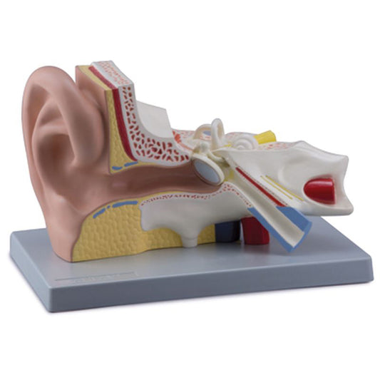
Classic ear model which has been enlarged
Regular price €102,95 EURRegular priceUnit price per -
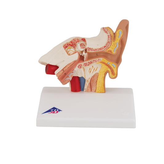
Very enlarged and detailed ear model which can be separated into 4 parts
Regular price €133,95 EURRegular priceUnit price per -
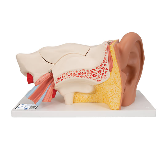
Ear model 3 times enlarged - can be separated into 5 parts
Regular price €231,95 EURRegular priceUnit price per -
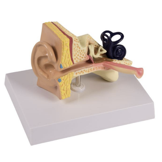
Ear model of a child's ear showing otitis media
Regular price €102,95 EURRegular priceUnit price per -
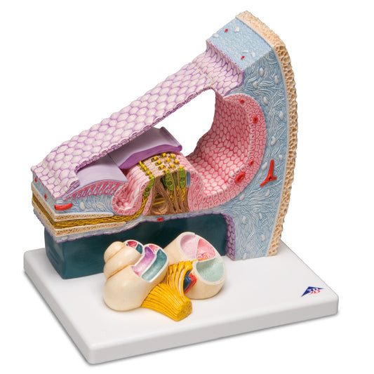
Detailed ear model that shows both the entire cochlea and a cross-section with three-dimensional details
Regular price €324,95 EURRegular priceUnit price per -
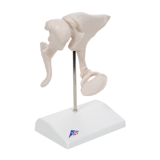
Life-size model of the 3 small bones of the middle ear
Regular price €140,95 EURRegular priceUnit price per -
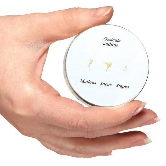
20 x magnification of the bones in the middle ear (ossicula auditus)
Regular price €233,95 EURRegular priceUnit price per -
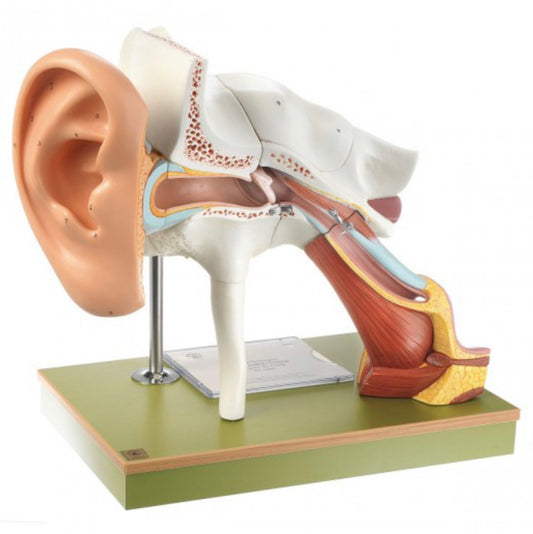
Enlarged ear model with auricula of the highest quality
Regular price €1.750,95 EURRegular priceUnit price per -
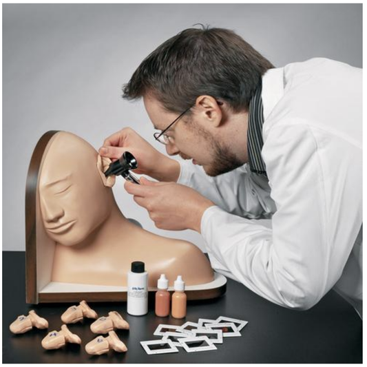
Simulator for training in ear examination
Regular price €2.047,95 EURRegular priceUnit price per

A window to a world of anatomy
Whatever you're looking for
Then we can procure or produce it. eAnatomi is more than just a retailer of existing products. We have our own development department, where we create unique and original products that are used for training, guidance and inspiration.

19 years of anatomy
A safe transaction
For 19 years I have been at the head of eAnatomi and sold anatomical models and posters to 'almost everyone' working with anatomy in Denmark and abroad. When you do business with eAnatomi, you do business with me and I personally guarantee a safe transaction.
Christian Birksø
Owner and founder of eAnatomi
Latest blog news
View all-
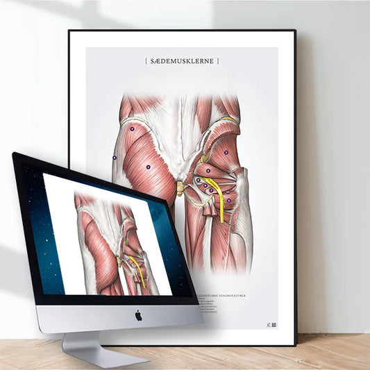
Digital anatomical illustrations
With eAnatomi and our new offer of digital anatomical illustrations, you can now legally acquire a license to upgrade your digital communication, regardless of whether it is for educational use,...
Digital anatomical illustrations
With eAnatomi and our new offer of digital anatomical illustrations, you can now legally acquire a license to upgrade your digital communication, regardless of whether it is for educational use,...
-

Anatomical Chart Company - changes the format
Anatomical Chart Company har i 2023 besluttet af udfase papirvarianten for fremover kun at levere den let laminerede version med ringhuller. Dette betyder at det ikke længere bliver muligt, at...
Anatomical Chart Company - changes the format
Anatomical Chart Company har i 2023 besluttet af udfase papirvarianten for fremover kun at levere den let laminerede version med ringhuller. Dette betyder at det ikke længere bliver muligt, at...
-

The heart model's place in teaching & expla...
Teachers, professionals and other mediators can easily be challenged when they have to explain the anatomy and diseases of the heart.
The heart model's place in teaching & expla...
Teachers, professionals and other mediators can easily be challenged when they have to explain the anatomy and diseases of the heart.


